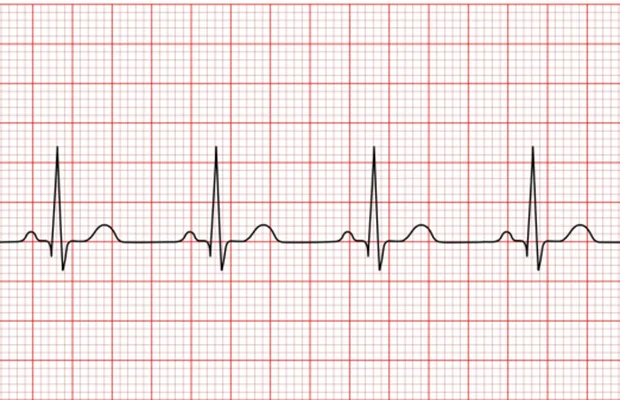
What’s an Electrocardiogram (ECG)?

An ECG is a simple non-invasive test in which electrodes are placed on the chest, arms and legs to record the electrical activity of the heart. It takes less than 10 minutes and no preparation is required. It gives the cardiologist valuable information about your heart rhythm. 10 electrodes are placed on your chest and attached to the monitor. The ECG machine records 10 seconds of your heart’s electrical activity. The heart’s contraction is represented by wave forms on the ECG which are interpreted by the cardiologist.

How the ECG Works: A Simple Overview

  • Heart contraction: The heart’s contraction can be seen on the ECG. A normal ECG shows sinus rhythm and is made up of a series of waves called the P, QRS and T wave.
  • Atrial contraction: Atrial activation is represented by the ‘P-Wave’ on the ECG.
  • Ventricular contraction: Ventricular activation is represented by the ‘QRS’ complex on the ECG. This normally takes less than 120 milliseconds. When the Ventricles are activated, blood is pumped from the heart to the lungs and through the aorta to the rest of the body.
  • Ventricular relaxation: The final wave is called the T wave. During this phase the Ventricles relax. This normally takes less than 450 milliseconds.

Why is the ECG Important?

The ECG gives a quick overview of the electrical signal in the heart. It helps spot anything unusual, from rhythm disturbances to more significant concerns. The ECG can also be used by the cardiologist with concern to prescribing certain medications. Changes to an individuals ECG can have important clinical relevance, therefore we routinely perform ECGs for comparison with a patient’s baseline ECG.

ENQUIRE NOW

Fill out the form below and we will get back to you as soon as possible.